Today ultrasound is routinely used throughout pregnancy, but it is still relatively a new development in obstetrics. The first use of ultrasounds during pregnancy occurred in the late 1950s in Scotland, but it wasn’t until well into the 1970s that ultrasound during pregnancy became routine in the US.
Ultrasound transformed our experience of pregnancy
Prior to the introduction of home pregnancy tests and obstetric ultrasound in the 1970s, pregnancy was a rather mysterious affair. Women had to guess if they were pregnant based on a lack of periods and their symptoms, and in order to confirm the pregnancy they had to go to a doctor for a pregnancy test; thus, many women were not sure if they were pregnant until well into the first trimester or possibly into the 2nd trimester.
Once the pregnancy was confirmed, the woman just kind of waited until the baby was born and everyone hoped for the best. Many of the pieces of information modern couples think of as normal were a complete mystery back then: an accurate due date, the sex of the fetus, whether there was more than one fetus, and in particular, whether the fetus was developing normally. Prior to ultrasound, it was a “wait and hope” experience. Nowadays, many couples get multiple ultrasounds throughout the pregnancy and know every detail about the fetus’s development, the exact due date, and as the third trimester progresses, are usually quite confident several months before birth as to the number of babies they will have, the sex of the baby, and that the baby will be healthy.
The many uses of ultrasound
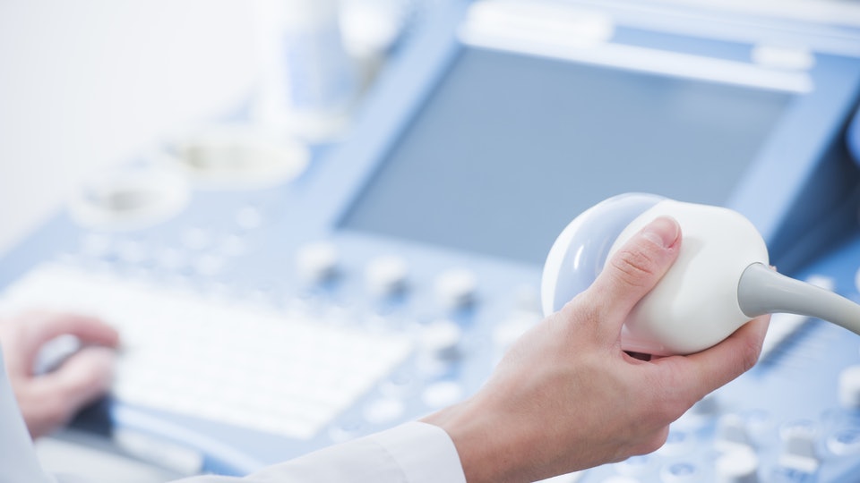
Ultrasound has become a routine tool in the obstetrician’s office. The ultrasound machine cost, even for really advanced machines that provide 3D images, is generally considered to be well worth it by most doctors.
Ectopic pregnancies
Currently, women are urged to seek an ultrasound as soon as they confirm they are pregnant, which for some women is as early as 6 to 8 weeks. The embryo at this stage is tiny and barely visible, and the primary purpose of this examination is to a) confirm the pregnancy, and to b) confirm the pregnancy is not ectopic, namely the embryo has implanted inside the uterus instead of somewhere else. Prior to widespread use of early ultrasounds, women didn’t know they had an ectopic pregnancy until they were in the process of bleeding to death.
First trimester screening for major birth defects
Women are urged to undergo another ultrasound around 12 weeks of gestation, and the primary purpose of this examination is to detect major malfunctions of fetal development, including serious heart defects, spina bifida, and chromosomal number aberrations like trisomy 13 and 21. This screening examination offers prospective parents the chance to quickly terminate a severely abnormal fetus instead of the only option in the past of not knowing and being blindsided by a stillbirth, a severely malformed baby that died shortly after birth, or a severely malformed baby that will never be able to live a normal life.
Second trimester screening for birth defects
Another ultrasound is generally recommended around 18 to 20 weeks to perform a more thorough examination of the fetus’s health. At this stage, the fetus is large enough that most abnormalities of development become glaringly obvious. The state of the internal organs can be evaluated, the number of fingers and toes can be counted, and the sex of the fetus can be determined. If something is seriously awry, again this screening offers the opportunity to terminate, or, for most couples, it offers reassurance that their baby will be born healthy.
Third trimester ultrasounds
Third trimester ultrasounds are usually only conducted if something is going not quite right with the pregnancy; these screenings have saved many a mother’s and baby’s life by allowing the doctor to quickly determine that the baby needs to come out right now to save mother and baby. Prior to ultrasound, people were simply surprised when these horrible events occurred.
Confirming a miscarriage/stillbirth
Unfortunately, miscarriages, particularly early in pregnancy, are quite common. Without ultrasound, miscarrying women were unable to determine exactly what was happening-was the fetus dead for sure? And the doctors could not determine if the entire fetus and placenta were expelled; retained tissue can kill the recently-pregnant woman, but with ultrasound, obtaining exact details about what is going wrong is relatively easy.
In summary
In summary, obstetric ultrasound has totally transformed our experience of pregnancy. It used to be a mysterious, potentially dangerous experience that could rapidly kill a woman at various stages; an event that potentially led to birth being a stressful event since no one knew what might emerge from the womb until it did. Now, with ultrasound, every detail of the developmental process can be followed throughout the pregnancy, leading to much greater safety for the mother and very few surprises occurring after birth. Although some may mourn the loss of the “mystery”, most couples and doctors much prefer knowing as much as possible.


